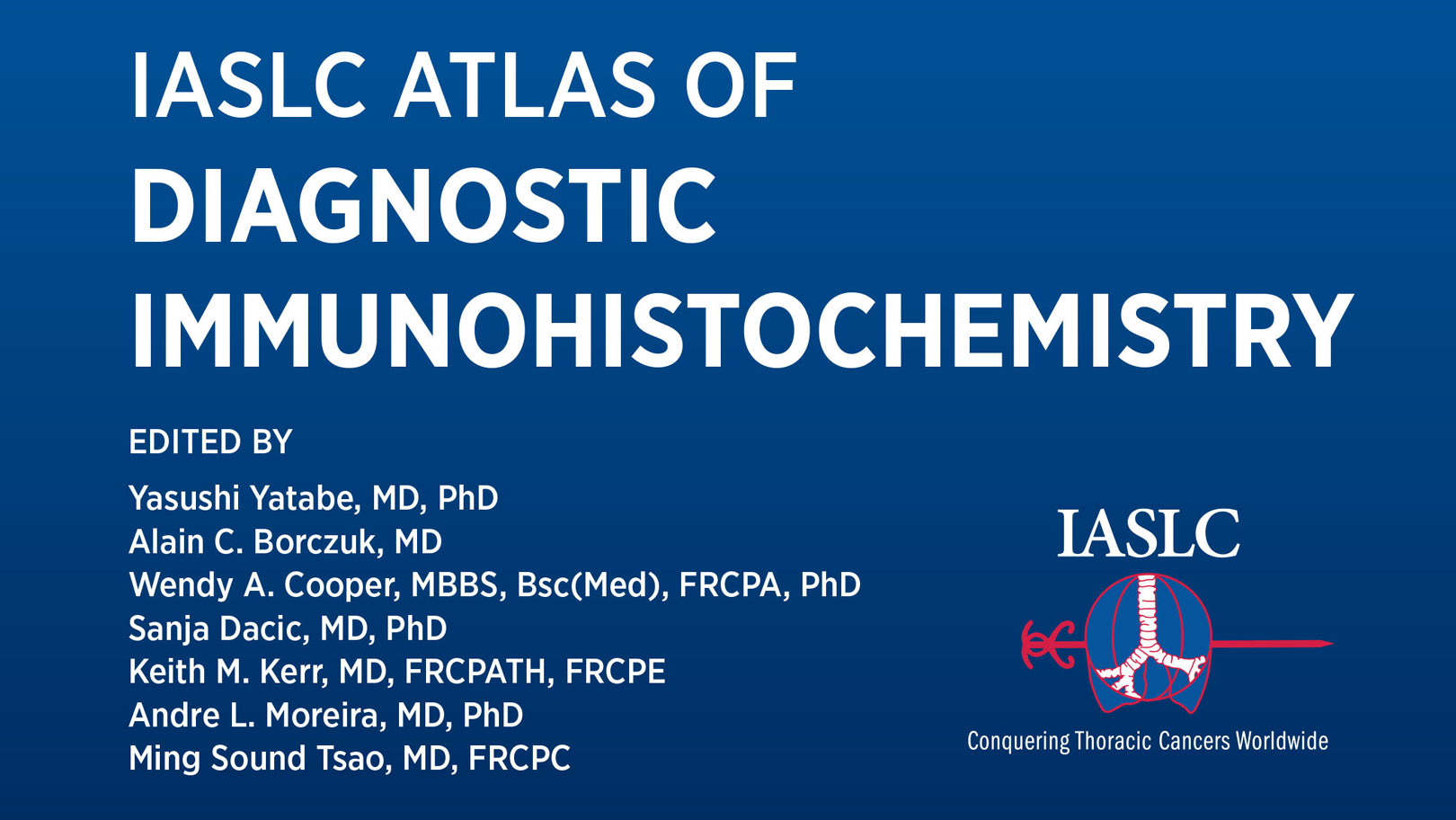
The latest in the series of diagnostic Atlases has been published by the IASLC and its Pathology Committee. Following the highly successful editions on ALK, EGFR, ROS1 and PD-L1, this latest Atlas takes a comprehensive look at diagnostic immunohistochemistry (IHC) and its role in thoracic pathology. The editorial team of Drs. Yasushi Yatabe, Alain Borczuk, Wendy Cooper, Sanja Dacic, Keith Kerr, Andre Moreira, and Ming Tsao brought together a series of short chapters written by members of the Pathology Committee and other IASLC members to give a practical perspective of this enormously important diagnostic technique.
In 2018, the IASLC Pathology Committee published best practice recommendations for diagnostic immunohistochemistry in thoracic malignancies in the Journal of Thoracic Oncology.1 Because of the nature of a review article, the recommendations had to be concise, and only a limited number of figures were included. After the article achieved significant popularity, the committee members realized that there was more to offer and began work on the Atlas. The chapters in this volume are largely built around a series of key questions and answers, but also featuring more discursive text and copious colour illustrations. IHC is a fundamental technique used in the diagnosis of many thoracic malignancies and this Atlas provides, in its twenty chapters and addenda, a succinct, well-illustrated, contemporary discussion of the key elements in a reader-friendly format. It provides not only a practical commentary for pathologists, but also a clinical perspective on how important IHC is in innumerable aspects of thoracic tumor diagnosis.
Practical and Detailed: Understanding the Role for IHC
Introductory perspectives from the editors, all practicing thoracic pathologists, are followed by a clinical take on diagnostic IHC by Drs. Harvey Pass and Balazs Halmos. Several generic chapters on techniques and technical issues in IHC then follow, providing important information on basic elements of how these assays work, how and when they should be used, and how they should be interpreted. Also included are traditional discussions of IHC expression in all types of thoracic tumour from the most common to the most rare, as well as several chapters dedicated to the most common markers used in lung cancer diagnosis. Immunohistochemical expression of some markers has become a defining feature in the classification of common lung cancer subtypes described in the WHO classification—the ‘rule book’ that most pathologists worldwide use to diagnose and classify surgically resected lung and thoracic tumors. IHC has also become a crucial diagnostic adjunct in the accurate diagnosis and classification of tumor samples taken during small biopsy procedures and by cytologic techniques; this Atlas provides a complementary guide to practice, providing concise, useful explanations as well as more in-depth detailed discussion of this vital, everyday scenario. IHC has transformed our ability to accurately diagnose and subtype thoracic cancers, including mesothelioma and thymoma. Judicious use of IHC underpins the subclassification of NSCLC samples with implications for both chemotherapy choice and triage of cases for predictive biomarker testing, when traditional morphology alone is not sufficient.
IHC Helps Guide Therapy Choice
In this era of personalized medicine for patients with thoracic malignancy, and NSCLC in particular, underpinned by a greater understanding of the molecular basis of neoplasia and routine genotyping of clinical tumor samples to inform patients treatment choice, it might seem something of a throwback that a technique that has been in the pathology laboratory for 40 years is still being used. But used it should be! On the one hand, keen observers of contemporary pathology practice could be forgiven for thinking that today’s pathologists never make any diagnosis without the aid of IHC. This is not, and should not be, true. Basic morphologic assessment using tinctorial tissue stains is still the basis of pathologic diagnosis. But IHC can provide, when required, crucial supportive evidence of a suspected diagnosis, or a completely new perspective on the nature of tumor cells based on protein expression—evidence that can completely alter the final diagnosis and improve diagnostic accuracy. IHC marker expression may have quantitative as well as qualitative elements, and can also provide a spatial perspective relating protein expression to cell type and location within the tumor or within individual tumor cells. Once these factors become better understood and gain clinical relevance, the result is better classification of disease, better diagnosis and ultimately, better therapy choices for patients. IHC has an important role in predictive biomarker testing for therapy targets in patients with NSCLC, some of which was described in the ALK/ROS1 and PD-L1 Atlases. Although genomic alterations form the basis of those tumors that may respond to a number of such targeted therapies, it is the resultant abnormal (onco)protein that exerts an oncologic effect, and which is the target of the drug. It thus makes complete sense that assessment of that protein could have importance in patient stratification.
The editors, and the team at IASLC headquarters led by Dr. Jill Daigneault who worked on this Atlas, are immensely grateful to myriad colleagues and friends who gave up their time to contribute their deep and wide-ranging expertise to author this book. We are also indebted to the many sponsors from industry who provided much needed support to allow for the production of this title. Particularly, we were encouraged that the extent of this support reaffirmed an understanding across the oncology community of the crucial importance of these first steps in pathologic diagnosis . We are sure that this Atlas will be a useful reference for anyone interested in thoracic tumor diagnostics, from basic researchers to practising oncologists.
Reference:
- Yatabe Y, Dacic S, Borczuk AC, et al for the IASLC Pathology Committee. Best Practices Recommendations for Diagnostic Immunohistochemistry in Lung Cancer. J Thorac Oncol. 2018;14(3):337-407.


